Digital radiography has become a cornerstone of modern dental care, giving patients clearer images, faster results, and a gentler diagnostic experience. At the practice of Hearth Dental Practice, we rely on digital sensors and advanced imaging software to capture detailed views of teeth, roots, and supporting bone—information that helps us make timely, well-informed decisions about your oral health. This page explains what digital radiography is, how it works, and why it matters for your dental care.
In simple terms, digital radiography replaces traditional film with electronic sensors that capture X-ray images and transfer them immediately to a computer. The sensor detects X-ray energy and converts it into a digital image that the dentist can manipulate—zooming, adjusting contrast, and measuring structures without waiting for film processing. For patients, this means the same diagnostic capability as film radiographs but with a more streamlined workflow and clearer visual information.
Digital images are stored as data rather than physical prints, which makes them easy to archive, retrieve, and review during follow-up appointments. Because they can be enhanced on-screen, clinicians often spot subtle changes earlier than they might on conventional film. That early detection supports more conservative treatment plans and interventions that focus on preserving natural tooth structure.
While “digital radiography” covers a few different sensor types and technologies, the clinical goal is consistent: to produce high-quality images that support accurate diagnosis, efficient communication, and precise treatment planning. The result is better-informed care that aligns with modern standards for safety and effectiveness.
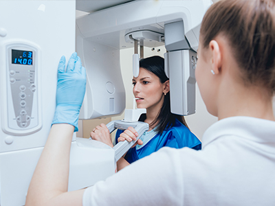
Digital sensors come in a variety of shapes and sizes, designed to fit comfortably in the mouth and capture specific regions—bitewing sensors for interproximal views, periapical sensors for individual teeth, and panoramic systems for broader overviews. Sensors are usually housed in thin, flexible casings that minimize gag reflexes and improve patient comfort compared with older film holders.
The imaging process itself is quick. A brief exposure produces the image, and within seconds the photograph appears on the monitor. Because exposures are shorter and the equipment is more efficient, patients typically spend less time in the chair during the imaging portion of an appointment. Clinicians can confirm image quality immediately and retake a shot only if necessary, reducing inconvenience for patients.
Clinicians also use positioning aids and gentle technique to make the experience as comfortable as possible. If you have concerns about sensitivity or anxiety during X-rays, your dental team will explain each step and accommodate special needs to ensure a calm, safe imaging session.
One of the most important benefits of digital radiography is reduced radiation exposure compared with traditional film radiographs. Digital sensors are more sensitive to X-rays, which allows for shorter exposure times while still producing diagnostic-quality images. This sensitivity, combined with modern X-ray generators that focus the beam precisely, helps minimize the overall radiation dose for patients.
Beyond lower exposure, digital systems offer improved control over image acquisition and evaluation. Enhanced image processing can reveal diagnostic detail that might otherwise require additional shots, so clinicians often gather the necessary information with fewer exposures. Proper training and adherence to safety protocols ensure that radiographs continue to be used judiciously and only when they benefit patient care.
Pregnancy and pediatric care are situations where imaging decisions are made with extra care; when radiographs are necessary, the team follows established guidelines to prioritize safety—using shielding, limiting the number of images, and selecting the lowest acceptable exposure settings while maintaining image quality.
Because digital images are available instantly, dental teams can review findings with patients during the same visit. This immediacy improves communication: clinicians can point out areas of concern, annotate images, and compare current images with past records to track changes. Visual feedback helps patients understand their condition and participate more effectively in treatment decisions.
Digital radiographs also integrate seamlessly with treatment planning tools and electronic health records. Images can be calibrated and measured precisely, aiding in restorative work, implant planning, and periodontal assessments. When combined with other digital tools, such as intraoral scanners, radiography contributes to a coordinated digital workflow that supports predictable, minimally invasive care.
In multi-disciplinary cases, digital files can be shared quickly and securely with specialists for collaborative planning. This efficient exchange reduces delays and ensures that all members of a patient’s care team are working from the same accurate, high-resolution images.
Digital radiography eliminates the need for chemical processing and physical film storage, which reduces environmental waste and simplifies office logistics. There’s no darkroom or developing chemicals to handle, and digital archives free up physical space while improving accessibility. For practices, this translates to a smaller environmental footprint and more streamlined administrative processes.
Storing images electronically also improves recordkeeping and continuity of care. Digital files are indexed and backed up, reducing the risk of lost images and making it easier to retrieve historical data for long-term monitoring. When patients move or change providers, electronic images can be transferred efficiently—helping maintain continuity while preserving diagnostic history.
Additionally, digital systems support robust data security practices when configured correctly. Access controls, encrypted backups, and secure transfer protocols help protect patient information while enabling convenient clinical use. This combination of environmental and technical advantages makes digital radiography a practical, forward-looking choice for modern dental care.
Digital radiography is a practical innovation that enhances clinical insight, patient safety, and office efficiency. By combining sensitive sensors, instant imaging, and integrated software, this technology supports conservative, well-informed dental care. If you have questions about how digital imaging will be used in your treatment, or if you’re interested in seeing sample images and a brief explanation during your visit, please contact the practice to learn more. We welcome the opportunity to explain how digital radiography contributes to precise, patient-centered care at Hearth Dental Practice


Digital radiography is an X-ray imaging method that uses electronic sensors instead of traditional photographic film to capture detailed images of teeth, roots and supporting bone. The sensor converts X-ray energy into a digital image that appears on a computer monitor within seconds, allowing immediate review and manipulation. This rapid availability helps clinicians detect concerns earlier and plan care more efficiently.
Because images are saved as digital data rather than physical prints, they are easy to archive, retrieve and compare over time. Enhanced image processing lets clinicians adjust brightness, contrast and magnification to reveal subtle changes that might be missed on film. The result is clearer diagnostic information that supports conservative, evidence-based treatment decisions.
Digital radiography replaces photographic film with sensitive electronic sensors that require much shorter exposure times to produce diagnostic images. Images appear instantly on a computer, eliminating the need for chemical processing and reducing the overall imaging workflow. The increased sensitivity and immediate feedback typically reduce the need for repeat exposures.
Digital images can be enhanced, measured and annotated on-screen, which improves diagnostic clarity and clinician-patient communication. The ability to calibrate and compare images precisely supports restorative planning and long-term monitoring. In practice, these capabilities make imaging faster, more flexible and often more informative than film-based systems.
Digital radiography generally exposes patients to lower levels of radiation than traditional film X-rays because electronic sensors are more efficient at capturing X-ray photons. Modern X-ray generators and collimation techniques further limit the beam to the area of interest, minimizing scatter and unnecessary exposure. Clinicians follow established safety protocols to ensure exposures are as low as reasonably achievable while still obtaining diagnostic images.
Special considerations are applied for pregnant patients and children, including using shielding and limiting the number of images to what is clinically necessary. Your dental team will discuss any concerns and tailor imaging to your individual needs so that safety remains a top priority. If additional precautions are needed, the team will explain them before imaging begins.
Sensors come in a variety of sizes for bitewing, periapical and panoramic imaging and are enclosed in thin, biocompatible housings to improve comfort. During the exposure, the sensor is positioned in the mouth with gentle positioning aids and the X-ray source is briefly activated, producing an image in a fraction of a second. Most patients describe the procedure as quick and no more uncomfortable than holding a small object in place for a short time.
Because images appear instantly, clinicians can confirm image quality immediately and repeat a shot only if necessary, reducing overall time in the chair. Technicians use careful technique and patient communication to minimize gag reflex and anxiety. If you have special needs or sensitivity concerns, let the team know so they can accommodate you comfortably.
Digital radiographs provide high-resolution visual information that clinicians use to detect decay, evaluate root structure, assess bone levels and plan restorative or surgical treatments. Images can be annotated and compared side-by-side with prior studies, which helps track changes over time and supports earlier, more conservative interventions. Instant availability lets clinicians review findings with patients during the same visit to improve understanding and shared decision-making.
Digital files integrate with treatment-planning tools and electronic health records, enabling precise measurements for implant placement, crown preparation and periodontal assessment. In complex cases, these images can be securely shared with specialists to coordinate multidisciplinary care. This seamless workflow promotes predictable outcomes and reduces the need for redundant imaging.
By revealing small lesions and early structural changes, digital radiography helps clinicians intervene at an earlier stage when treatments can be less invasive and more focused on preserving natural tooth structure. Enhanced visualization and precise measurements guide conservative restorations, targeted preventive measures and minimally invasive surgical planning. This aligns with a philosophy of preserving healthy tissue and promoting natural remineralization whenever possible.
When combined with other digital tools like intraoral scanners and clinical photography, radiographs contribute to a coordinated diagnostic picture that reduces guesswork. Early detection and accurate imaging often mean smaller restorations, fewer appointments and longer-lasting outcomes. The net effect is care that emphasizes preservation, comfort and long-term oral health.
Yes, digital radiographs can be exported and shared with other clinicians in standard file formats so that specialists and referral providers can review them quickly. Electronic sharing significantly reduces the time required for collaborative case planning and helps ensure that all members of a patient’s care team are working from the same high-quality images. Transfers are typically faster and more efficient than mailing physical film or prints.
When images are shared, the practice follows secure transfer protocols and privacy safeguards to protect your health information. Your dental team will obtain any necessary consents and confirm recipient contact details before sending files. This approach balances timely consultation with adherence to data security and confidentiality standards.
Digital radiographs are stored as indexed files within the practice’s imaging system and electronic health record, which makes retrieval simple and reliable for follow-up care. Regular backups and organized archiving reduce the risk of lost records and support long-term monitoring of dental health. Digital storage also frees the office from maintaining physical film archives and chemical processing supplies.
Access to stored images is controlled by user permissions and secure authentication, and responsible practices employ encrypted backups and secure transfer methods. These measures help protect patient privacy while maintaining clinical accessibility. If you have questions about how your records are stored or how to request a copy, the dental team can explain their specific procedures.
The frequency of dental X-rays varies depending on your individual oral health, risk factors, symptoms and whether you are a new patient needing a baseline set of images. Rather than following a one-size-fits-all schedule, clinicians tailor imaging intervals to each patient to ensure appropriate monitoring while minimizing unnecessary exposure. Factors such as recent decay, periodontal disease, ongoing restorative work or developmental concerns influence how often images are recommended.
Your dentist will review your medical and dental history, conduct an examination and discuss the rationale for any recommended radiographs. Because digital systems make it easier to compare current and past studies, they support targeted monitoring strategies over time. If you prefer more information about the recommended imaging plan, ask your clinician to explain how it relates to your specific needs.
At your appointment, the team will explain the imaging process, position a small sensor or use an external panoramic unit depending on the images needed, and take one or more exposures that appear on the monitor within seconds. The clinician will review the images with you, pointing out findings and describing any recommended follow-up or treatment options. The entire procedure is typically brief and designed to be as comfortable as possible for patients of all ages.
Safety measures such as thyroid shielding and careful technique are used when appropriate, and special precautions are taken for pregnant patients and children to minimize exposure. If you have anxiety, sensitivity or mobility concerns, let the staff know and they will accommodate your needs. For patients visiting the Mountain View office, the team is available to answer questions and demonstrate images to help you understand your oral health.
Ready to schedule your next dental appointment or have questions about our services?
Contacting Hearth Dental Practice is easy! Our friendly staff is available to assist you with scheduling appointments, answering inquiries about treatment options, and addressing any concerns you may have. Whether you prefer to give us a call, send us an email, or fill out our convenient online contact form, we're here to help. Don't wait to take the first step towards achieving the smile of your dreams – reach out to us today and discover the difference personalized dental care can make.