An intraoral camera is a compact, pen-sized imaging device designed to capture high-resolution color images from inside the mouth. Equipped with a small lens and built-in lighting, it brings a magnified, close-up perspective of teeth, gums, and other oral tissues that you can view on a computer monitor in real time. Unlike traditional mirrors, the camera provides a steady, illuminated view that helps spot subtle surface changes and hidden areas that are difficult to see with the naked eye.
These cameras record crisp stills and short video clips that reflect natural color and texture, making it easier to distinguish fractures, wear patterns, stain, and early signs of gum inflammation. Their ergonomic design lets clinicians maneuver gently around the mouth for comprehensive imaging—front and back teeth, beneath restorations, and along the gumline—without causing discomfort. The result is a level of visual detail that supports accurate assessment and clear communication.
Modern intraoral cameras are reliable, safe, and simple to use. They connect directly to the office computer or imaging system and integrate with electronic health records, streamlining how visual information is saved and retrieved. For patients, the experience is noninvasive and quick; for clinicians, it provides a practical diagnostic tool that complements other assessments like visual exams and radiographs.
One of the most important roles an intraoral camera plays is improving communication between clinician and patient. Instead of relying on verbal descriptions or small mirror views, dentists can show patients a magnified, full-color image of a specific tooth or area of concern. Seeing the issue directly helps patients understand what the clinician observes, why a recommendation is being made, and how different treatment options compare.
This visual approach supports informed decision-making by reducing ambiguity. When patients can see the exact location and appearance of a problem—such as a hairline crack, early decay, or a failing margin around a restoration—they are better positioned to ask targeted questions and participate in treatment planning. It also helps clarify the purpose and expected outcome of recommended procedures, which builds trust and reduces anxiety.
Because intraoral images are instantaneous, they can drive productive in-chair conversations. Clinicians can annotate, zoom, and compare images side-by-side to illustrate progression or improvement over time. That immediacy turns a routine examination into an interactive educational moment, reinforcing preventive guidance and making follow-up care easier to understand and remember.
Captured images from intraoral cameras become part of the patient’s permanent record, providing a visual baseline that supports diagnosis, monitoring, and legal documentation. These images are invaluable for tracking changes across visits—whether monitoring a lesion, assessing the progression of wear, or confirming the integrity of a restored tooth. Stored alongside clinical notes and radiographs, they add a layer of objective evidence to each patient chart.
In many cases, intraoral photographs are used to coordinate care with specialists and dental laboratories. High-quality images help implant surgeons, orthodontists, endodontists, and lab technicians understand the clinical situation without relying solely on written descriptions. When a clinician shares images with a laboratory, for example, technicians can better match shade, shape, and margin details, which improves the final restoration’s fit and appearance.
Images can also support administrative processes when required—such as referrals or documentation submitted for review—while preserving patient confidentiality within secure systems. Because the photos are reproducible and standardized, they streamline collaboration and reduce the back-and-forth that can delay treatment planning.
Intraoral cameras align closely with conservative dentistry principles by enabling earlier detection and less invasive interventions. By revealing small lesions, hairline cracks, or early erosive changes that might otherwise go unnoticed, clinicians can apply preventive strategies such as remineralization, sealants, or targeted monitoring instead of immediately resorting to extensive restorations. The visual evidence helps prioritize preservation of healthy tooth structure.
When used alongside other digital tools—like digital radiography, intraoral scanners, and laser diagnostics—these cameras contribute to a comprehensive, technology-supported workflow. The combined data from multiple imaging modalities improves diagnostic confidence and supports precise, minimally disruptive treatments. This integrated approach reflects a broader commitment to durability and long-term oral health rather than quick fixes.
For practices focused on patient comfort and longevity of outcomes, intraoral imaging is a practical tool to make conservative care both understandable and achievable. It helps clinicians tailor interventions to the actual state of the tissues while documenting the rationale for less invasive options, reinforcing a treatment philosophy centered on preservation and prevention.
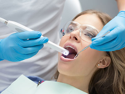
A typical intraoral camera exam is quick, comfortable, and noninvasive. During a routine checkup or when evaluating a specific concern, the clinician will gently guide the camera into the mouth, positioning it to capture the areas of interest. Lighting and magnification are adjusted automatically or via the imaging software, and the patient watches the images appear on the screen. The entire process usually takes only a few minutes and requires no special preparation.
Because the camera is small and portable, it can reach difficult angles with minimal discomfort, even for patients who are sensitive to long appointments or gag reflexes. The images are saved to the patient’s file immediately, so clinicians can review them again later or include them in follow-up discussions. If necessary, clinicians may take several angles to ensure a comprehensive visual record of the area in question.
Safety and hygiene are priorities: the device is cleaned and sterilized between uses or covered with appropriate disposable sleeves, depending on the design. Patients who wish to keep copies of their images for personal reference can request them through normal office procedures, and clinicians will use the images to illustrate preventive recommendations and any planned treatments.
Overall, intraoral camera exams enhance transparency and understanding while remaining a simple, routine part of comprehensive dental care. By making oral conditions visible and measurable, they support clearer communication, more conservative decision-making, and better long-term outcomes for patients.
Summary
An intraoral camera is a practical, patient-friendly imaging tool that brings detailed, real-time views of the mouth to life. It strengthens communication, improves clinical documentation, and supports minimally invasive treatment planning. In the office of Hearth Dental Practice, this technology is used to help patients understand their oral health and make informed decisions with confidence. Contact us to learn more about how intraoral imaging may be used in your care.


An intraoral camera is a small, pen-sized digital imaging device used to photograph the inside of the mouth. It combines magnification, built-in lighting and a miniature lens to reveal details of teeth, gums and oral tissues that are difficult to see with the naked eye. Images and short video clips appear in real time on a monitor so clinicians and patients can review findings together.
These cameras produce high-resolution stills that preserve natural color and surface texture, which helps distinguish fractures, wear patterns and early signs of decay or inflammation. Most modern units connect to the office computer and integrate with electronic health records for efficient storage. The device is noninvasive and designed for gentle intraoral navigation to minimize discomfort during imaging.
An intraoral camera turns abstract descriptions into clear visual evidence by showing patients a magnified, full-color image of the specific area of concern. When patients see the condition directly—such as a hairline crack or early decay—they can ask targeted questions and better understand why a clinician recommends particular options. This transparency reduces ambiguity and supports informed decision-making.
Clinicians can annotate, zoom and compare images side by side to illustrate progression or results of previous treatment, which makes explanations more concrete. Instant imaging also allows for productive in-chair conversations during the exam rather than relying solely on verbal summaries later. The visual approach builds understanding and helps patients remember preventive guidance and follow-up instructions.
An intraoral camera helps reveal a wide range of conditions that may be hard to spot with a mirror alone, including early enamel lesions, hairline fractures, worn restorations and localized gum inflammation. The magnified, high-resolution images make subtle surface changes and stain patterns more visible, which supports early intervention. In some cases the camera can show areas beneath or adjacent to restorations that warrant closer evaluation.
While intraoral imaging complements radiographs and tactile exams, it is particularly valuable for documenting surface details and soft-tissue appearance. Images can aid in monitoring lesions, assessing margin integrity of crowns and fillings, and identifying areas of unusual wear or erosion. For comprehensive diagnosis, clinicians combine camera images with other diagnostic tools to form a complete clinical picture.
Yes. Intraoral cameras are noninvasive, use no ionizing radiation and are designed to be comfortable for patients of all ages. The devices are small and maneuverable, allowing clinicians to reach difficult angles without causing significant gagging or extended discomfort. Lighting and focus are adjusted through the imaging software, so exposure is limited to visible light only.
Clinics follow strict infection-control protocols by cleaning and sterilizing the device between uses or by using disposable sleeves where appropriate. The quick nature of the exam—typically only a few minutes for targeted imaging—reduces chair time for sensitive patients. If patients want copies of their images for personal reference, offices can provide them through normal record-request procedures while maintaining privacy safeguards.
Intraoral photographs are saved directly to the patient’s electronic chart and become part of the permanent record, providing a visual baseline for diagnosis and monitoring. Because images are date-stamped and reproducible, they are useful for tracking changes over time and documenting the condition of tissues before and after treatment. These records add objective evidence that complements written notes and radiographs.
Stored images also support administrative tasks like referrals and clinical documentation without compromising patient confidentiality when managed through secure systems. Clinicians can retrieve prior images to compare progression or healing, which improves continuity of care. High-quality photographs also help standardize communication when discussing cases with specialists or laboratory technicians.
Intraoral imaging advances conservative dentistry by enabling earlier detection of small lesions, cracks and erosive changes that might otherwise go unnoticed. Seeing subtle problems sooner allows clinicians to recommend preventive or minimally invasive interventions—such as remineralization strategies, sealants or targeted monitoring—instead of immediately performing extensive restorations. The visual evidence helps justify a preservation-first approach to treatment planning.
When combined with other digital tools like digital radiography and intraoral scanners, camera images contribute to a comprehensive diagnostic workflow that increases diagnostic confidence. This multimodal data supports precise, smaller interventions tailored to the tooth’s actual condition and helps avoid unnecessary removal of healthy tooth structure. In the office of Angela Laithangbam, DDS Inc., intraoral imaging is one component of a technology-supported approach that prioritizes long-term oral health.
High-quality intraoral images are a practical way to convey clinical details to specialists and dental laboratories without relying solely on written descriptions. Photographs can illustrate shade, shape, margin details and the relationship of restorations to surrounding tissues, which helps implant surgeons, orthodontists, endodontists and lab technicians understand the clinical situation more clearly. This visual information reduces the need for repeated clarification and streamlines case planning.
When sharing images, clinicians use secure, HIPAA-compliant systems to protect patient privacy while ensuring collaborators have the information they need. Technicians can use the photos to match esthetic details more accurately and to fabricate restorations that fit both functionally and visually. The result is more efficient coordination and improved predictability of outcomes.
An intraoral camera exam is typically quick and straightforward, often performed as part of a routine checkup or when evaluating a specific concern. The clinician will gently guide the camera to the area of interest while you watch the images appear on a monitor, and lighting and focus are adjusted through the software. Multiple angles may be captured to create a comprehensive visual record of the site in question.
The process usually takes only a few minutes and requires no special preparation from the patient. Devices are handled with strict hygiene practices, including sterilization or disposable sleeves between uses. After the exam the clinician will review and explain the images, and they may save selected photos to your chart for future comparison.
Absolutely. Because intraoral images are reproducible and stored in the patient record with dates, they make it easy to compare the condition of teeth and soft tissues across visits. Clinicians can track progression of lesions, changes in restoration margins, or healing after treatment by reviewing sequential photographs. Side-by-side comparisons help determine whether a conservative approach is effective or whether further intervention is needed.
Regular photographic monitoring is particularly useful for early lesions, cracks and areas of wear where visual change can guide timing of treatment. Image archives also provide objective documentation that supports long-term maintenance plans and patient education. This documented timeline enhances continuity of care and improves diagnostic transparency.
At Angela Laithangbam, DDS Inc., intraoral imaging is integrated into routine examinations and used as a communication and diagnostic aid to support conservative, patient-centered care. Images are used to explain findings during the appointment, document baseline conditions and coordinate care with specialists or laboratories when needed. The practice emphasizes minimally invasive strategies and uses imaging to identify problems early and tailor interventions accordingly.
Patients can expect clear explanations supported by visual evidence, and images are saved securely to the electronic health record for future reference. This approach aligns with the practice’s focus on gentle, technology-supported dentistry that prioritizes preservation and long-term oral health. If you have questions about how intraoral imaging may be applied in your care, the Mountain View office staff can discuss the process during your next visit.
Ready to schedule your next dental appointment or have questions about our services?
Contacting Hearth Dental Practice is easy! Our friendly staff is available to assist you with scheduling appointments, answering inquiries about treatment options, and addressing any concerns you may have. Whether you prefer to give us a call, send us an email, or fill out our convenient online contact form, we're here to help. Don't wait to take the first step towards achieving the smile of your dreams – reach out to us today and discover the difference personalized dental care can make.